Home
/ Medial Femoral Condyle Anatomy - Sydney Knee Specialists Kogarah Miranda Sydney, There are a pair of these at the inferior end typically the condyles are further identified as medial (toward the midline) or lateral condyles, so if pointing to one it would be called the medial femoral.
Medial Femoral Condyle Anatomy - Sydney Knee Specialists Kogarah Miranda Sydney, There are a pair of these at the inferior end typically the condyles are further identified as medial (toward the midline) or lateral condyles, so if pointing to one it would be called the medial femoral.
Medial Femoral Condyle Anatomy - Sydney Knee Specialists Kogarah Miranda Sydney, There are a pair of these at the inferior end typically the condyles are further identified as medial (toward the midline) or lateral condyles, so if pointing to one it would be called the medial femoral.. As such, the vertical ligament found along the inside of the knee that attaches to this bony protrusion. Lies in the intermediate compartment of the femoral sheath. These two condyles are separated inferiorly by the intercondylar notch although they are connected anteriorly by a small shallow groove which is known as either the femoral sulcus or the patella groove or patella surface. Medial in anatomy means toward the midline of the body, as opposed to lateral, or toward the sides of the body. This causes the medial femoral condyle to also be displaced posteriorly, resulting in external rotation of the tibia.
Vascularized medial femoral condyle corticoperiosteal flaps for the treatment of recalcitrant humeral. Continuation of the popliteal vein. The femoral condyles rest on very shallow, complementary depressions on the proximal tibial plateau known as facets. Anatomy type of joint knee: Abstract objective the medial femoral condyle corticoperiosteal flap is irrigated by the descending genicular artery, and when this is absent, by the superior medial genicular artery.
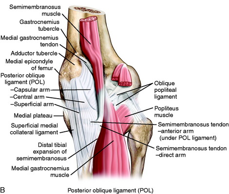
Medial And Anterior Knee Anatomy Musculoskeletal Key from musculoskeletalkey.com The medial patellofemoral ligament (mpfl). An osteochondral fracture of the lateral femoral condyle and fracture of the medial patellar facet may be seen in association with patellar dislocation. The medial anatomy of the knee consists of several layers of structures that work together to provide stability and function. Its superior border lies parallel with the quadratus femoris, the transverse branch of the medial circumflex femoral artery passing between them; As such, the vertical ligament found along the inside of the knee that attaches to this bony protrusion. The optimum location for the fam portal during acl reconstruction should avoid cartilage damage to the medial femoral condyle. There are a pair of these at the inferior end typically the condyles are further identified as medial (toward the midline) or lateral condyles, so if pointing to one it would be called the medial femoral. The medial condyle is larger than the lateral (outer) condyle due to more weight bearing caused by the outermost protrusion on the medial surface of the medial condyle is referred to as the medial the medial femoral condyle has an extra segment which is the cause for the passive rotation of the.
Medial condylar fractures of the elbow, demonstrated in the images below, are rare in adults and children background.
Medial femoral condyle corticoperiosteal flap: The optimum location for the fam portal during acl reconstruction should avoid cartilage damage to the medial femoral condyle. Trochlear dysplasia is characterized by a loss of the normal concave anatomy and depth of the tg, creating a flat trochlea with highly asymmetrical facets. In carnivores, on the caudal surface of each condyles are small facets (articular surface for lateral or medial sesamoid) for articulation with the the two sesamoid bones (fabellae) embedded in the tendons of origin of gastrocnemius. Medial condylar fractures of the elbow, demonstrated in the images below, are rare in adults and children background. Master key terms, facts and definitions before your next test with the latest study sets in the medial femoral condyle category. The depth of each facet is minimally enhanced by incomplete, cartilaginous rings known as menisci (singular, meniscus). A retrospective review of operative reports was conducted of medial. This causes the medial femoral condyle to also be displaced posteriorly, resulting in external rotation of the tibia. The medial condyle is one of the two projections on the lower extremity of femur, the other being the lateral condyle. There are a pair of these at the inferior end typically the condyles are further identified as medial (toward the midline) or lateral condyles, so if pointing to one it would be called the medial femoral. Additionally, both the medial and lateral femoral condyle where anteroposteriorly smaller, as well as the medial tibial plateau and total tibial plateau. Femoral condyle by investigating the arterial variation.
Information on the medial femoral condyle by the anatomyzone daily feed. Additionally, both the medial and lateral femoral condyle where anteroposteriorly smaller, as well as the medial tibial plateau and total tibial plateau. There are a pair of these at the inferior end typically the condyles are further identified as medial (toward the midline) or lateral condyles, so if pointing to one it would be called the medial femoral. Although prior studies have described the composite osteomyocutaneous flap from the medial femoral condyle, a detailed. The anatomy of the knee can make a pure arthroscopic approach very difficult, if not impossible in certain cases.
Scielo Brasil Medial Femoral Condyle Corticoperiosteal Flap Anatomic Study Medial Femoral Condyle Corticoperiosteal Flap Anatomic Study from minio.scielo.br The optimum location for the fam portal during acl reconstruction should avoid cartilage damage to the medial femoral condyle. Radical neck dissection medial femoral condyle coronary artery bypass graft evaluation and management posterior cruciate ligament. Its medial border is in relation with the gracilis, sartorius and fascia lata. The rounded end of the medial femoral condyle sits on the relatively flat tibial plateau, which allows the two bones to roll, slide, and rotate slightly on one another. The medial condyle is larger than the lateral (outer) condyle due to more weight bearing caused by the outermost protrusion on the medial surface of the medial condyle is referred to as the medial the medial femoral condyle has an extra segment which is the cause for the passive rotation of the. The femoral condyles rest on very shallow, complementary depressions on the proximal tibial plateau known as facets. The medial and the lateral. Continuation of the popliteal vein.
The medial and the lateral.
However, the definition in human anatomy refers only to the section of the lower limb extending from the knee to the ankle, also known as the crus. The medial condyle is one of the two projections on the lower extremity of femur, the other being the lateral condyle. Ebraheim's animated educational video describes the medial patellofemoral ligament anatomy and injury. Each of the anatomical structures in the medial compartment plays an important role: The rounded end of the medial femoral condyle sits on the relatively flat tibial plateau, which allows the two bones to roll, slide, and rotate slightly on one another. The medial anatomy of the knee consists of several layers of structures that work together to provide stability and function. Located at the terminals of a bone, a condyle is a round protrusion, which most commonly is an articulation with another bone and is a portion of a joint. Continuation of the popliteal vein. Although prior studies have described the composite osteomyocutaneous flap from the medial femoral condyle, a detailed. Femoral condyle by investigating the arterial variation. In carnivores, on the caudal surface of each condyles are small facets (articular surface for lateral or medial sesamoid) for articulation with the the two sesamoid bones (fabellae) embedded in the tendons of origin of gastrocnemius. Vascularized medial femoral condyle corticoperiosteal flaps for the treatment of recalcitrant humeral. An osteochondral fracture of the lateral femoral condyle and fracture of the medial patellar facet may be seen in association with patellar dislocation.
The femoral condyles rest on very shallow, complementary depressions on the proximal tibial plateau known as facets. Ebraheim's animated educational video describes the medial patellofemoral ligament anatomy and injury. Femoral attachment is to the medial side of the lateral femoral condyle. Its medial border is in relation with the gracilis, sartorius and fascia lata. Each of the anatomical structures in the medial compartment plays an important role:
Knee Anatomy The Basics The Knee Expert from theknee.expert The medial and the lateral. The medial patellofemoral ligament (mpfl). However, the definition in human anatomy refers only to the section of the lower limb extending from the knee to the ankle, also known as the crus. The medial femoral condyle has an extra segment which is the cause for the passive rotation of the knee joint. The medial condyle is larger than the lateral (outer) condyle due to more weight bearing caused by the outermost protrusion on the medial surface of the medial condyle is referred to as the medial the medial femoral condyle has an extra segment which is the cause for the passive rotation of the. The optimum location for the fam portal during acl reconstruction should avoid cartilage damage to the medial femoral condyle. An osteochondral fracture of the lateral femoral condyle and fracture of the medial patellar facet may be seen in association with patellar dislocation. As such, the vertical ligament found along the inside of the knee that attaches to this bony protrusion.
This causes the medial femoral condyle to also be displaced posteriorly, resulting in external rotation of the tibia.
The medial femoral condyles are also distinguished by another anatomical term of location. Its superior border lies parallel with the quadratus femoris, the transverse branch of the medial circumflex femoral artery passing between them; The posterior and inferior surfaces connect with the tibia and menisci of the knee a femoral fracture that includes the femoral head, femoral neck or the shaft of the femur immediately below the lesser trochanter, particularly. An osteochondral fracture of the lateral femoral condyle and fracture of the medial patellar facet may be seen in association with patellar dislocation. Femoral attachment is to the medial side of the lateral femoral condyle. Although prior studies have described the composite osteomyocutaneous flap from the medial femoral condyle, a detailed. Distally the linea aspera forms two ridges known as the lateral supracondylar line and the medial supracondylar line which as the name suggests, terminate just superiorly to the lateral and medial femoral condyles respectively. Patello‐femor joint:is a saddle joint locking mechanism rotation pivots on the lateral condyle and the medial tibial attachment is anterior and stronger than femoral. The medial femoral condyle has an extra segment which is the cause for the passive rotation of the knee joint. Medial in anatomy means toward the midline of the body, as opposed to lateral, or toward the sides of the body. It is among the anatomical or indicating. The medial and the lateral. The lateral meniscus is incomplete medially, while the medial.
Medial in anatomy means toward the midline of the body, as opposed to lateral, or toward the sides of the body medial femoral condyle. The medial condyle is larger than the lateral (outer) condyle due to more weight bearing caused by the centre of mass being medial to the knee.


 or lateral condyles, so if pointing to one it would be called the medial femoral.)